Advanced Eye Care Technology at Vani Vision
We harness the power of cutting-edge technology to enhance your vision and eye health. Our commitment to innovation drives us to offer advanced equipment and treatments, ensuring you receive the highest standard of eye care available today.
The Future of Eye Care: Our Solutions
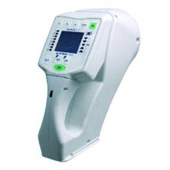
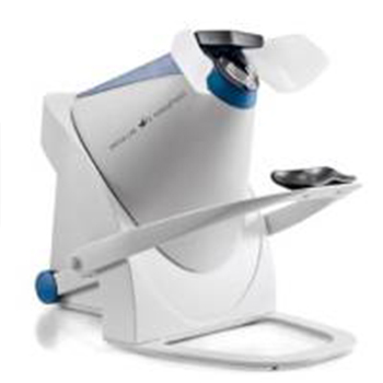
Corneal Topography with Advanced Topographer
Our topographer represents the pinnacle of corneal mapping technology. This sophisticated instrument is essential for accurately mapping the surface curvature of the cornea.
It is invaluable in fitting specialty contact lenses, especially for conditions like keratoconus. It ensures a precise fit for optimal vision correction and comfort.
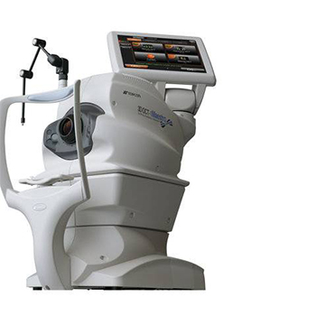
Retinal Health with OCT
The Optical Coherence Tomography (OCT) machine is a cornerstone of our retinal health monitoring arsenal. It provides us with high-resolution retina images, allowing for the early detection and ongoing monitoring of retinal diseases. This non-invasive imaging test is crucial for managing conditions such as age-related macular degeneration and diabetic retinopathy, helping to preserve vision.
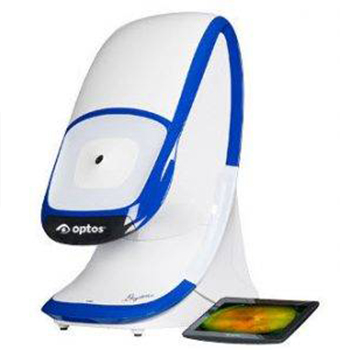
Wide-Field Retinal Imaging with Optomap Daytona
Our optomap Daytona retinal imaging system is a leap forward in retinal examination. It captures detailed, wide-field retina images, enabling us to assess your retinal health without dilation. This technology enhances patient comfort and allows for a more comprehensive view of the retinal surface, aiding in the early detection of eye conditions.
Visual Field Analysis with OCULUS Easyfield Perimeter
The OCULUS Easyfield perimeter offers precise and efficient visual field testing. This technology is particularly useful in diagnosing and monitoring glaucoma, allowing for early intervention. By mapping the visual field, the OCULUS Easyfield helps detect early signs of vision loss due to glaucoma, ensuring timely treatment to prevent progression.
Our adoption of these advanced technologies reflects our dedication to providing you with the best possible care. Whether it's fitting the perfect contact lens, monitoring retinal health, or managing glaucoma, we can meet your eye care needs with the highest standards of precision and care. Visit Vani Vision to experience the future of eye care, where innovative technology meets compassionate care.
- Phone: (770) 431-1713
- Fax: (470) 481 6736
- Email: info@vanivision.com
Atlanta, GA 30339
- Monday: 10:00 AM - 6:00 PM
- Tuesday: 10:00 AM - 6:00 PM
- Wednesday: 10:00 AM - 6:00 PM
- Thursday: 10:00 AM - 6:00 PM
- Friday: 10:00 AM - 5:00 PM
- Saturday: 9:30 AM - 1:30 PM
- Sunday: Closed